Here are some images that I've captured of my immunohistochemistry-stained brain tissue using a Leica Confocal TCS SP8 DLS microscope.

Parvalbumin interneurons (red), myelin (green), caspr (white) in the prefrontal cortex

Satellite oligodendrocytes (green) sitting in an unusual association with the parvalbumin (PV, blue) cell body and its associated perineuronal net (red). This image encapsulates the main finding of my PhD research, which is that the extracellular specializations surrounding PV undergo plasticity in a learning-dependent manner.

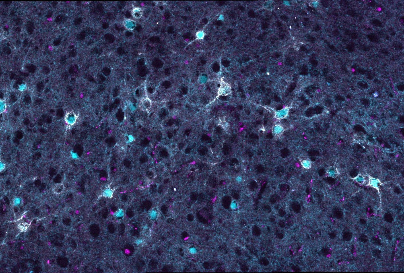
Parvalbumin interneurons (cyan) covered by perineuronal nets (white), interacting with oligodendrocyte lineage cells (magenta), in the nucleus accumbens

perineuronal nets in the nucleus accumbens

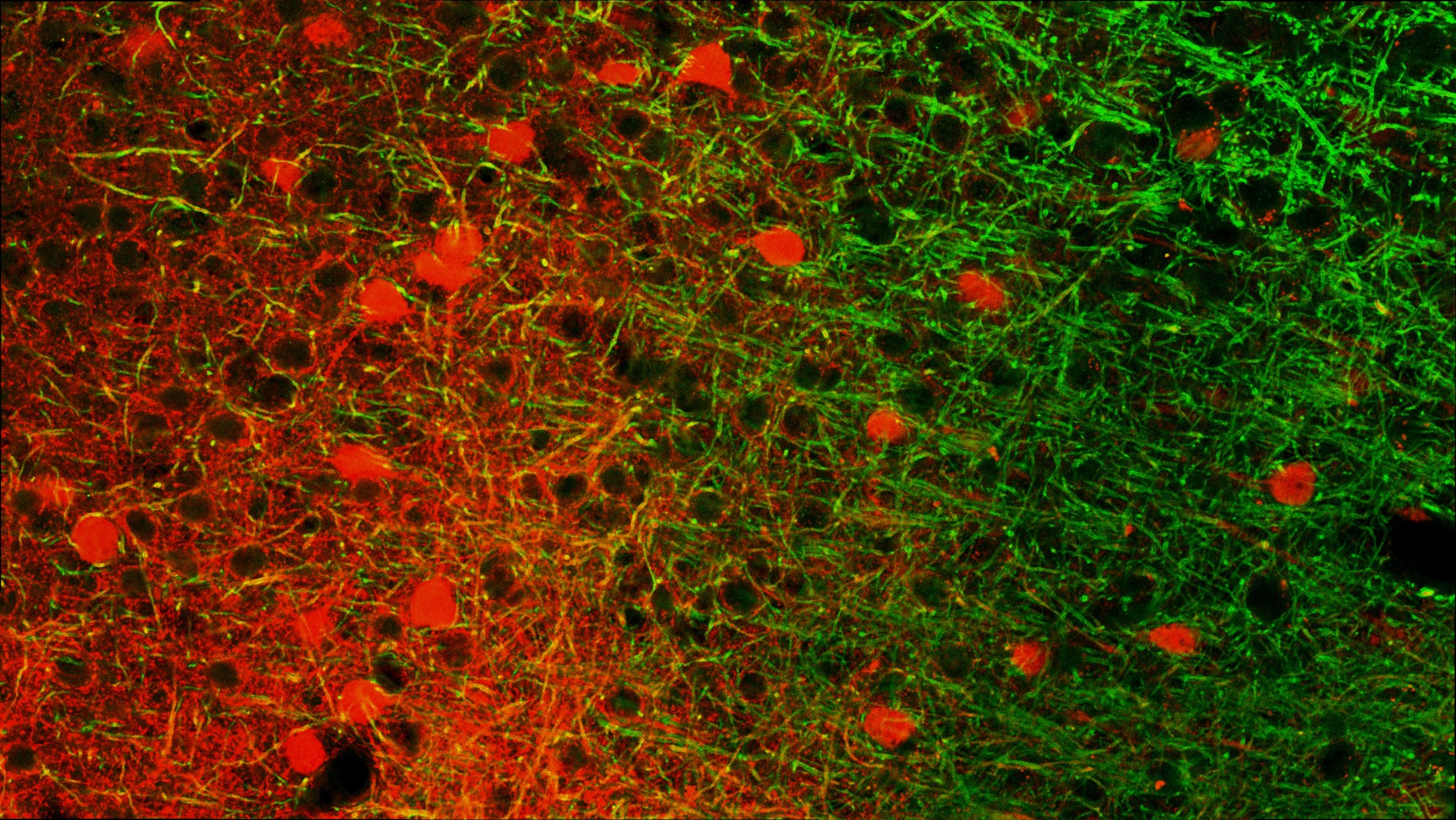
Myelinated (green) parvalbumin interneurons (red) in the prefrontal cortex

Parvalbumin interneurons (cyan) surrounded by perineuronal nets (white), interacting with oligodendrocytes (magenta) in the mouse cortex

A parvalbumin interneuron (red) dipping its axons into the heavily myelinated (green) prefrontal cortex

A rare sighting of a tiny myelinated segment of parvalbumin axon


